


3D Printed Right Ventricular Hypertrophy
Clinical History
This 56-year old female suffered from emphysema and gave a 2-year history of increasing
shortness of breath on exertion associated with recurrent attacks of bronchitis. On examination, she had a BP
160/90 mm Hg, pulse rate of 96 beats/min, and 6 cm of jugular venous congestion. The apex beat was impalpable,
bilateral crepitations were heard and pitting oedema was present peripherally. Special investigations: ECG
showed right heart strain pattern. Arterial blood examination showed respiratory acidosis. Despite treatment
there was steady deterioration and death.
Pathology
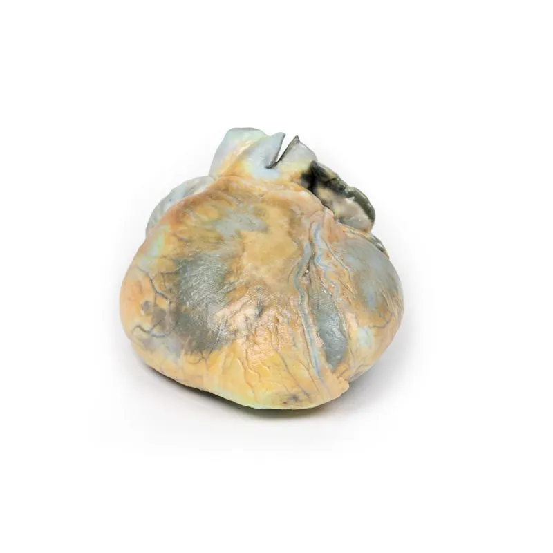
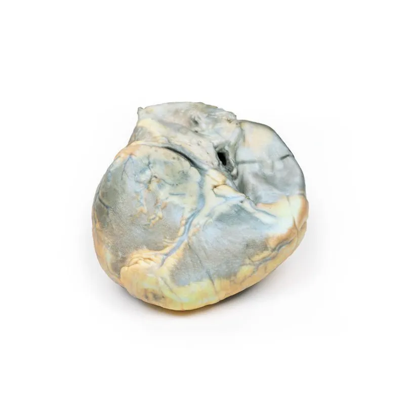
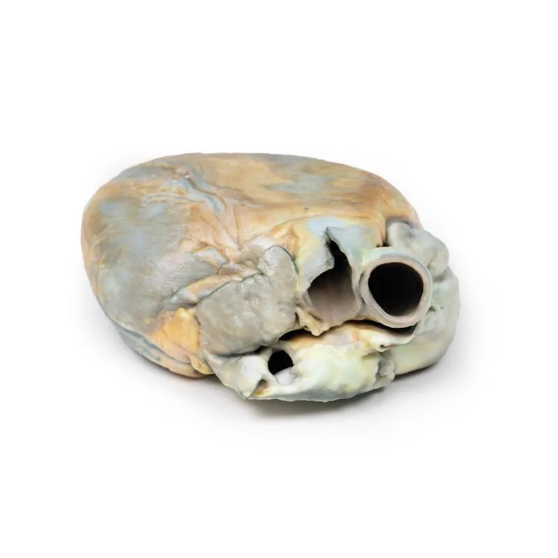
The specimen is of the external surface of the heart viewed from the anterior aspect. The right
ventricle is greatly enlarged and hypertrophied. All appears to be normal otherwise. This is an example of right
ventricular hypertrophy (RVH) in a patient with emphysema.
Further information
RVH usually occurs due to chronic lung disease or structural defects in the heart. One of
the most common causes of RVH is pulmonary hypertension (PH), which leads to increased pulmonary artery
pressure. As the right ventricle tries to compensate for this increased pressure it changes its shape and size
causing hypertrophy and right ventricular wall thickness. The global incidence of PH is 4 per 1M people: RVH
occurs in approximately 30% of these cases. Common causes of PH include chronic obstructive pulmonary disease
(COPD), pulmonary embolism, and other restrictive lung diseases. RVH also occurs in response to structural
defects in the heart, such as tricuspid insufficiency, which allows the backward flow of blood into the
ventricle. Other structural defects that lead to RVH include tetralogy of Fallot, ventricular septal defects,
pulmonary valve stenosis, and atrial septal defects. RVH is also associated with abdominal obesity and high
systolic blood pressure.
GTSimulators by Global Technologies
Erler Zimmer Authorized Dealer


